Ultrassom 360°
Learn to see beyond the image.
Explore anatomy, diagnosis, Doppler, musculoskeletal, POCUS, obstetrics, gynecology, pediatrics, guided procedures, and the new frontiers of ultrasonography.
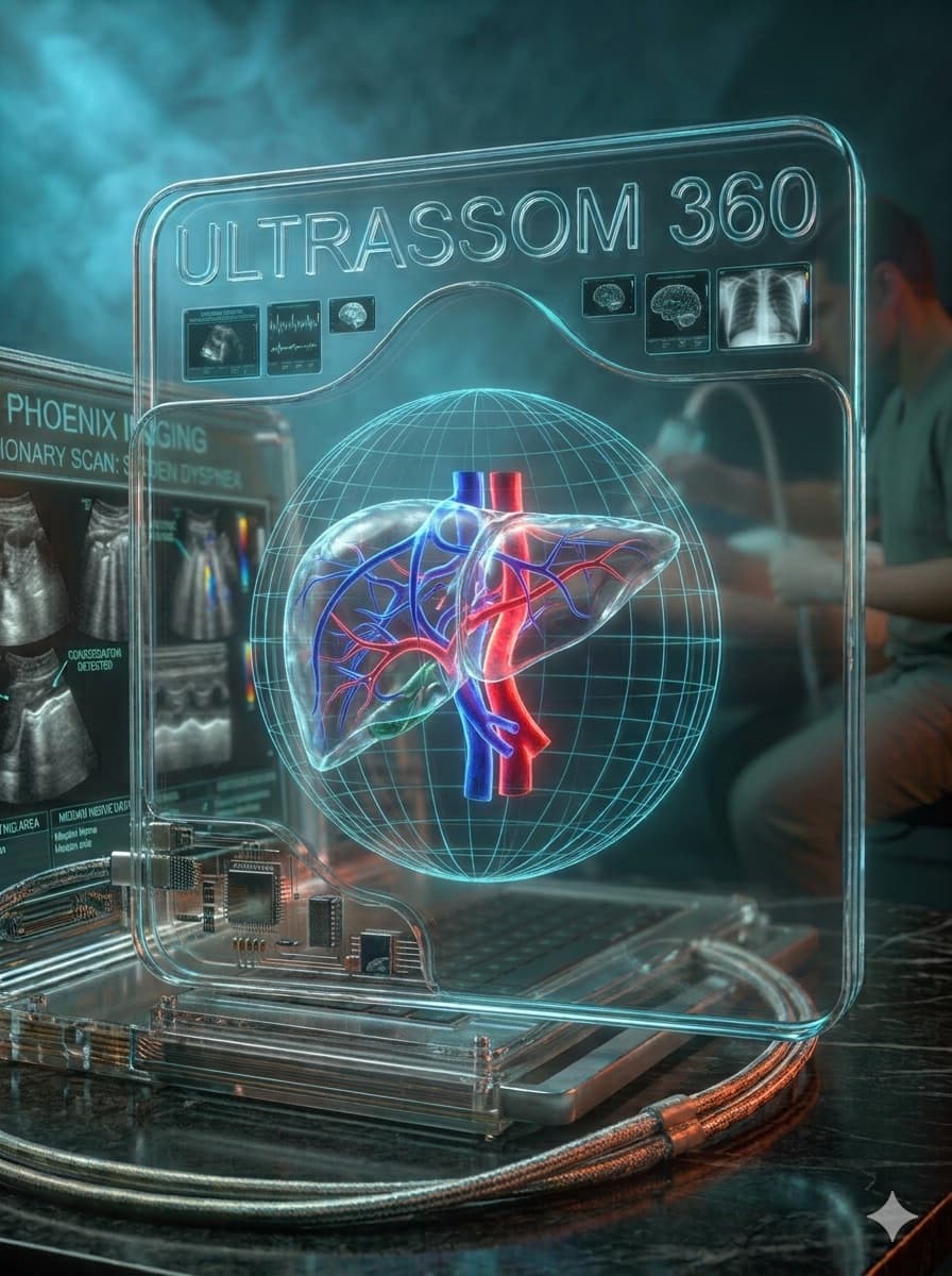
The 360° Method
Most courses teach you to press buttons on the machine. Here, every exam starts from the patient's complaint and ends in a clinical decision — not just a report.
Explore the Ultrasound Universe
Ultrasound goes far beyond the conventional abdominal exam — it's a broad, dynamic, portable diagnostic tool in constant evolution.
Musculoskeletal Ultrasound
Dynamic assessment of tendons, muscles, and joints — the fastest-growing modality among orthopedists, rheumatologists, and physiatrists.
Vascular Doppler
From arterial mapping to obstetric Doppler — blood flow as central diagnostic information.
Obstetric Ultrasonography
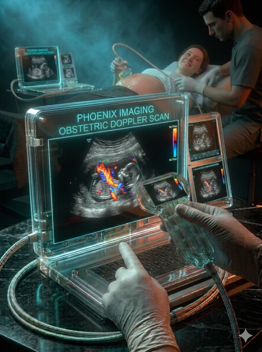
Pregnancy monitoring from the first to the third trimester, focused on biometry, morphology, and fetal well-being.
Gynecologic Ultrasonography
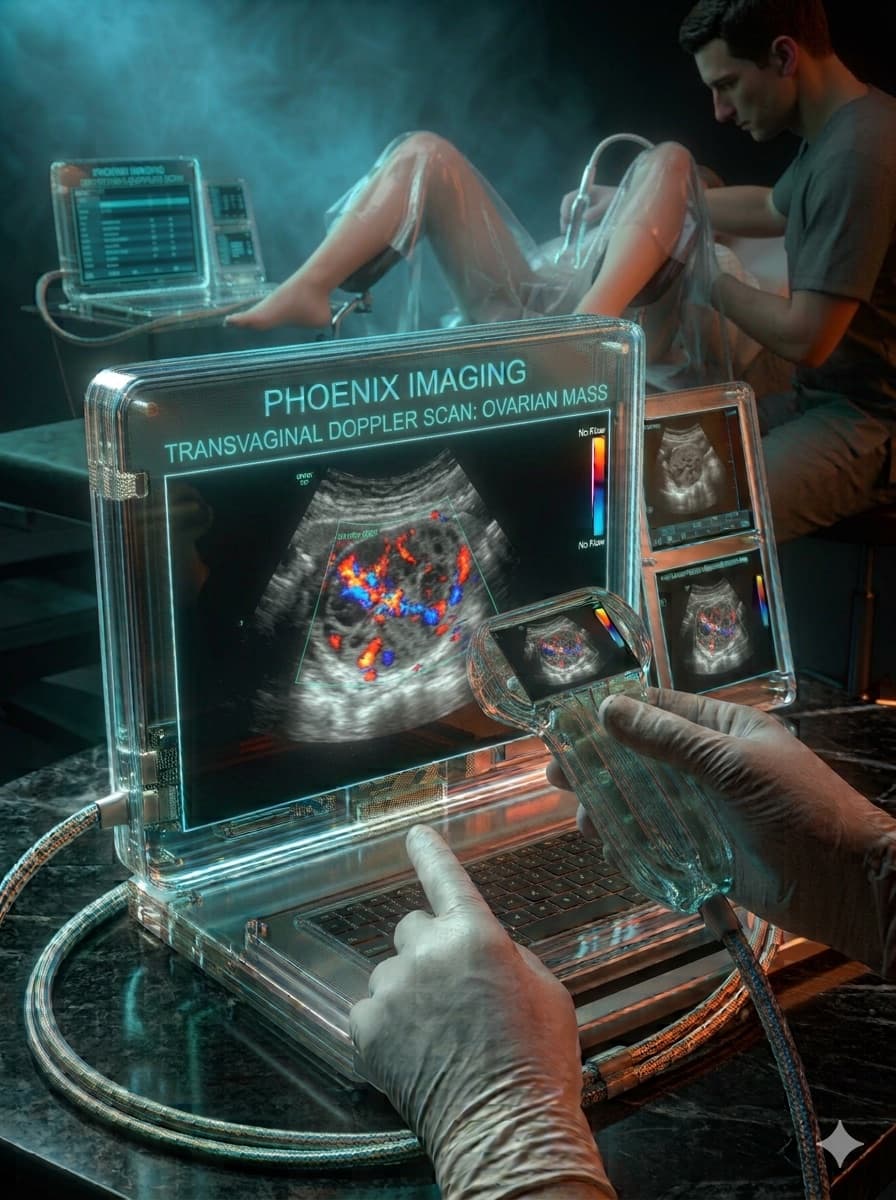
Uterus, ovaries, and adnexa investigated with transvaginal technique and Doppler, from routine exams to infertility workups.
Peripheral Nerve Ultrasound
Compressive neuropathies and neural anatomy mapped with high-resolution transducers.
Thoracic Ultrasound
Lung and pleura assessed at the bedside — one of the applications that most changed practice in emergency and critical care.
Head and Neck Ultrasound
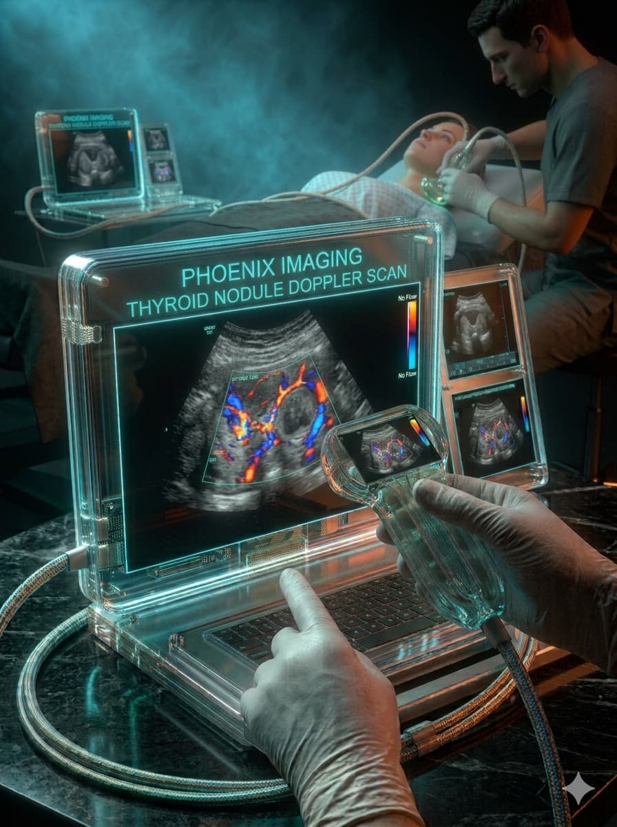
Thyroid, parathyroids, and lymph nodes — cervical anatomy of high clinical and surgical relevance.
Elastography
Tissue stiffness as diagnostic information — from the liver to the thyroid and breast.
Ultrassom 360° in action
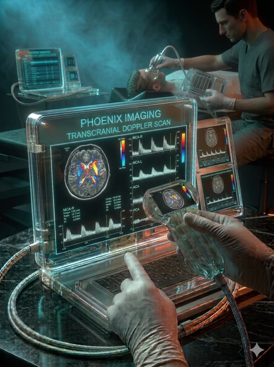
Real-time carotid Doppler, and a glimpse of findings spanning thyroid, obstetrics, gynecology, and neurology.
Real-time carotid vascular Doppler
Point-of-care ultrasonography
FAST, eFAST, pulmonary, cardiac, and shock assessment — the protocols that changed decision-making in urgent care, emergency, and critical care.
Learn about the protocolsTechnology in service of medical judgment
Pattern recognition, measurement automation, and image acquisition support. Artificial intelligence as a decision-support tool — never as a replacement for medical judgment.
See how AI is already usedClinical cases
Patient, indication, technique, findings, Doppler, and final diagnosis — in a format that helps you reason, not just memorize.
Incidental thyroid nodule — characterization and risk stratification
Solid hypoechoic nodule found on a routine exam, characterized and stratified using the TI-RADS system.
Ver caso completo →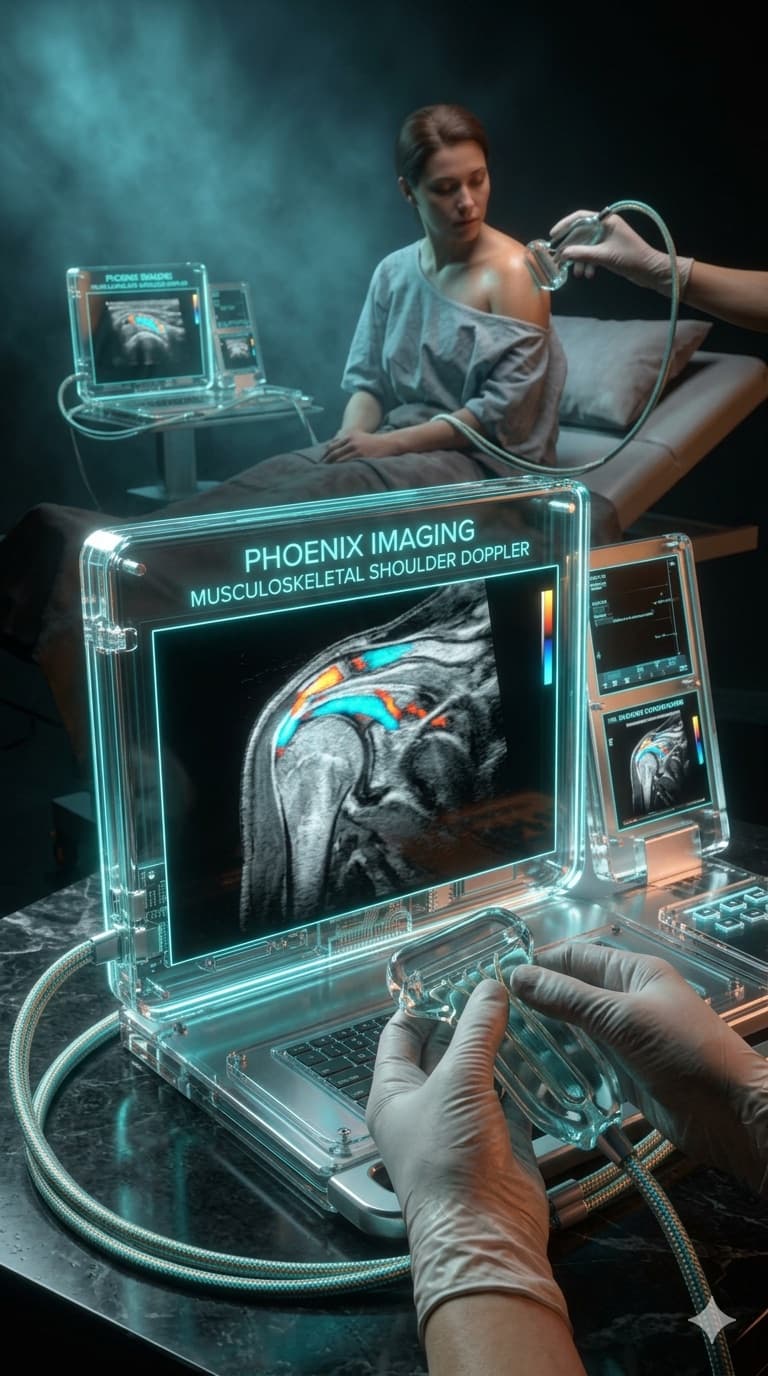
Shoulder pain in an athlete — dynamic assessment of the rotator cuff
Dynamic assessment of the supraspinatus tendon in an athlete with pain on abduction, with subacromial impingement documented in real time.
Ver caso completo →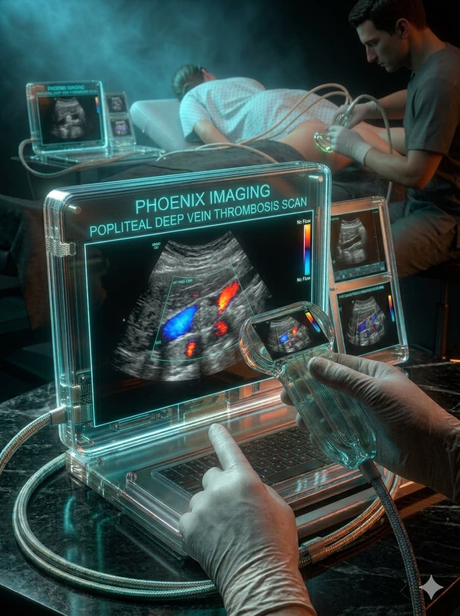
Unilateral lower limb edema — DVT workup
Lower limb venous Doppler in a patient with acute unilateral edema, confirming proximal deep vein thrombosis.
Ver caso completo →
MSK SmartClass: hands-on musculoskeletal ultrasound course
48 in-person hours, hands guided by Dr. Andrey Garcez — from normal sonographic anatomy to the most common pathologies, shoulder to foot.