Vascular Doppler
From arterial mapping to obstetric Doppler — blood flow as central diagnostic information.
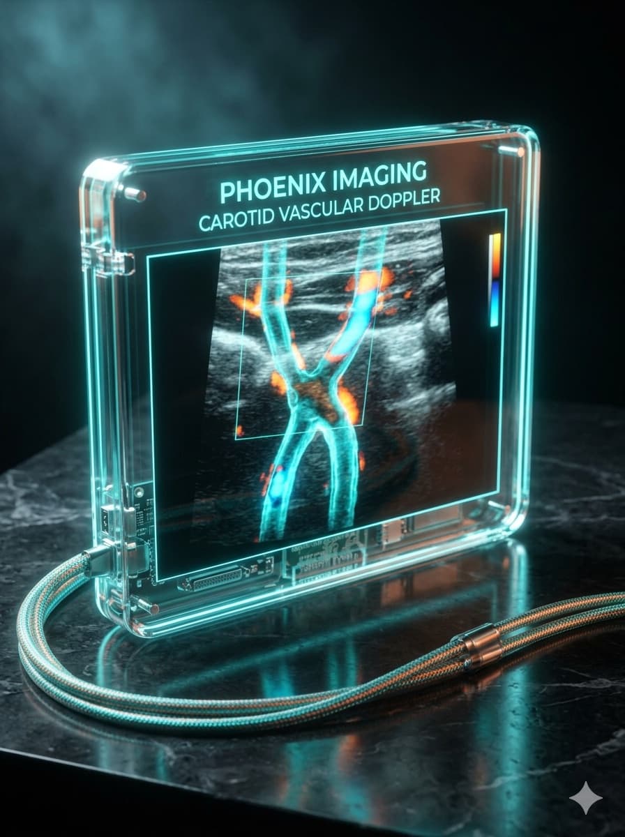
Doppler applications
Color Doppler
Maps flow direction and presence, essential for the rapid triage of vessels and structures.
Spectral Doppler
Quantifies velocities and waveform patterns — the basis for diagnostic criteria such as those for carotid stenosis.
Power Doppler
More sensitive to slow flow, useful in the vascularity of nodules and inflamed tissue.
In action
Carotid Doppler
Hepatic Doppler
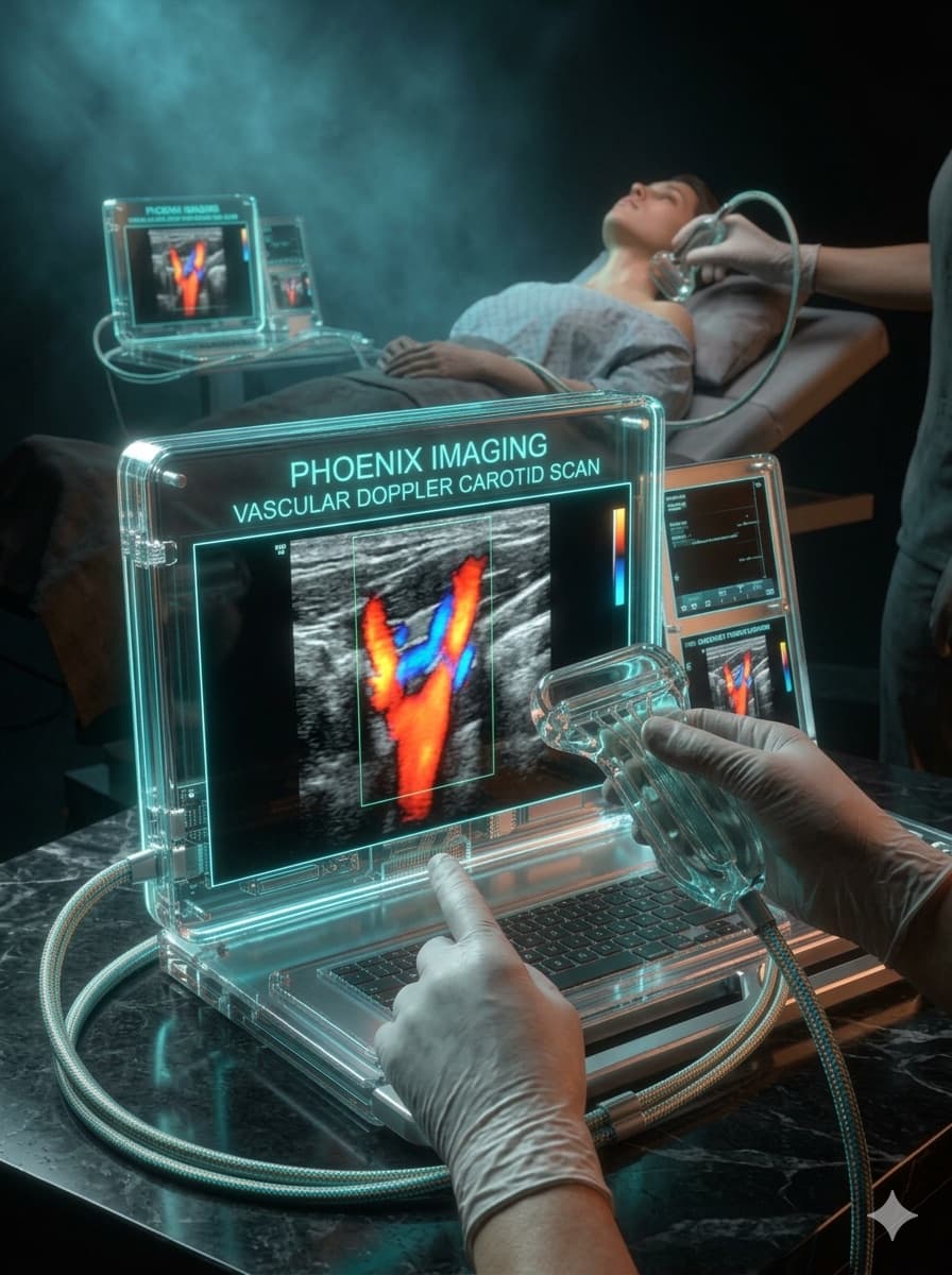
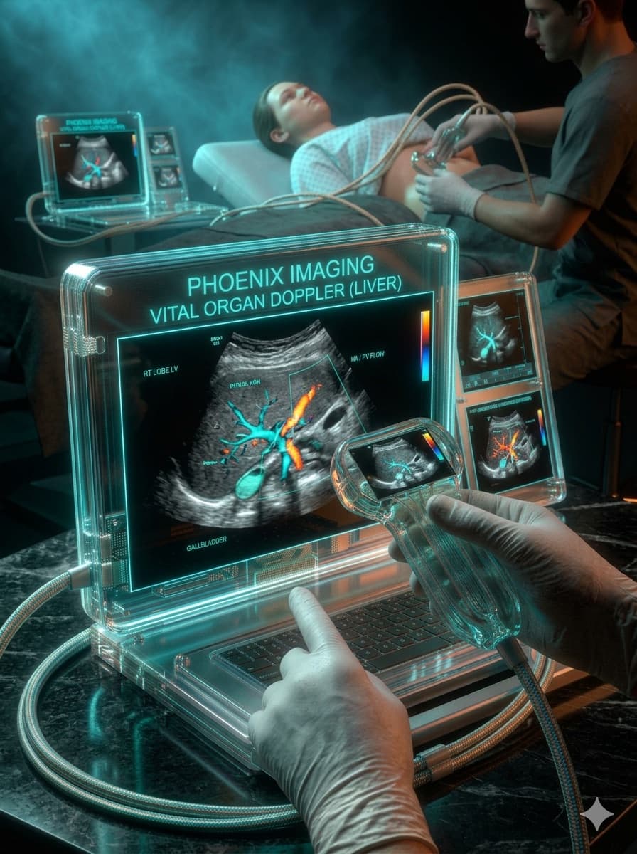
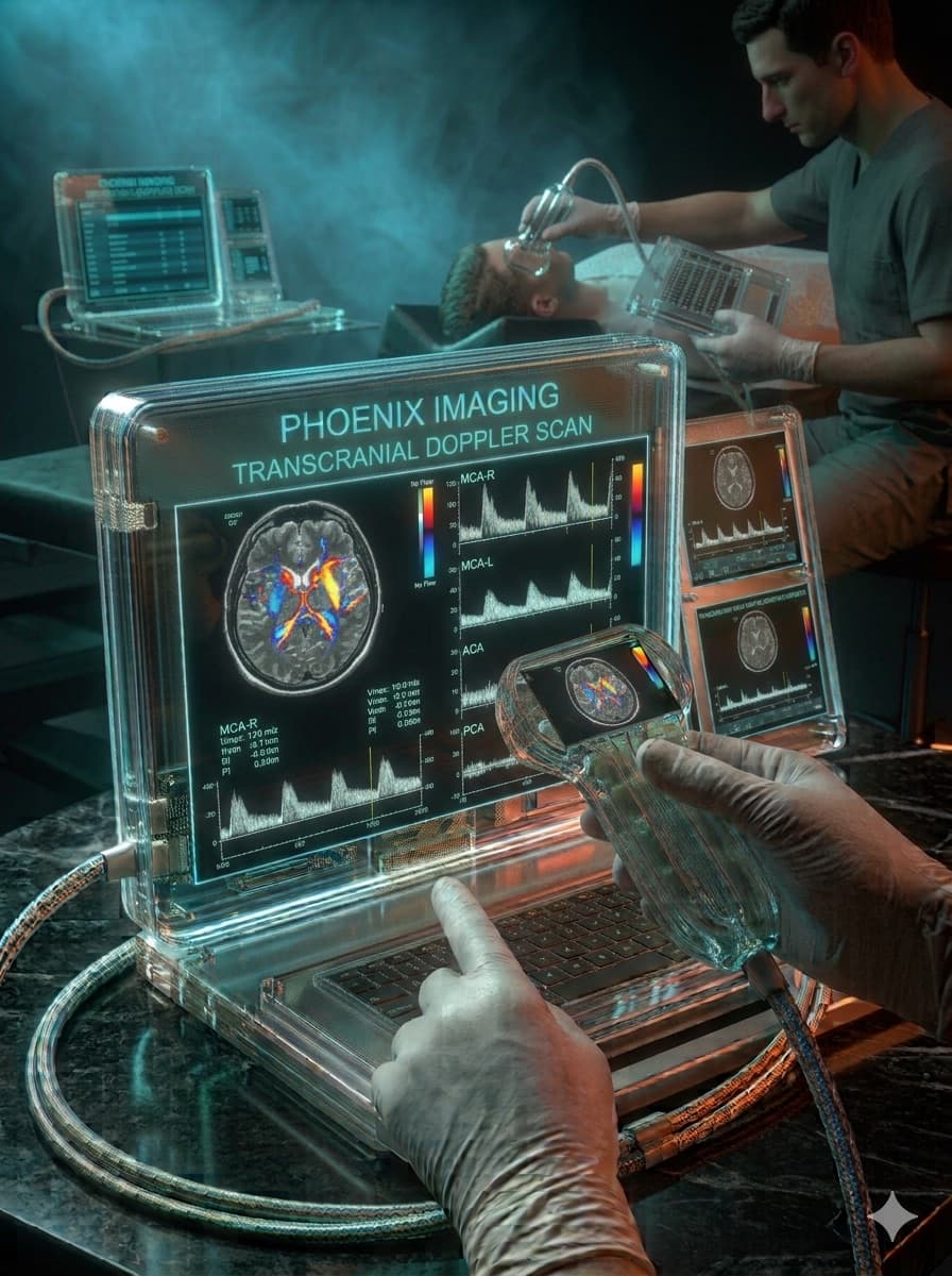

Doppler cases
See all →
Frequently asked questions
Is Doppler the same as a regular ultrasound?
No. B-mode ultrasound shows the anatomical structure (shape, size, texture of an organ or vessel), while Doppler adds blood flow assessment — direction, velocity and flow pattern inside veins and arteries. Both are used together in the same exam.
When does a doctor order a venous or arterial Doppler?
The most common indications include suspected deep vein thrombosis, investigation of arterial insufficiency (claudication, non-healing wounds), pre- and post-operative vascular assessment, and carotid plaque screening. The indication always starts from a clinical hypothesis.
Does a Doppler exam hurt or need special preparation?
It's not a painful exam. Abdominal Doppler usually requires 6 to 8 hours of fasting; limb or carotid Doppler usually needs no specific preparation — but it's worth confirming instructions with whoever ordered the exam.
What's the difference between color, spectral and power Doppler?
Color Doppler maps flow direction in colors overlaid on the image; spectral Doppler translates that flow into a velocity curve over time, essential for quantifying stenosis; power Doppler is more sensitive for detecting slow or low-volume flow, but without showing direction.
Want content like this before anyone else?
Join the Ultrassom 360° VIP list and get free lessons, commented clinical cases, and news before everyone else.