Unilateral lower limb edema — DVT workup
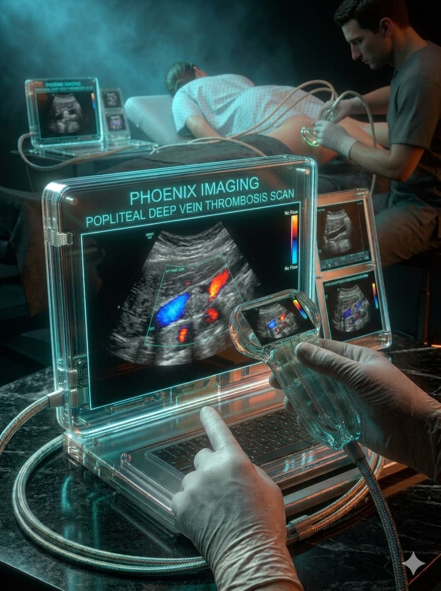
Patient, female, 61 years old, recent postoperative orthopedic surgery.
Unilateral edema and pain in the right lower limb, starting 48 hours ago.
Venous Doppler with serial compression of the deep venous system, from the femoral to the popliteal segment, combined with color and spectral Doppler.
Non-compressible common and superficial femoral veins, with echogenic endoluminal material and absent flow on color Doppler.
Absent spectral flow in the affected segments; phasic flow preserved in the non-thrombosed proximal and distal segments.
Acute deep vein thrombosis · Extrinsic venous compression · Ruptured Baker's cyst (clinical differential)
Acute deep vein thrombosis of the right common and superficial femoral veins.
Key points
- Venous non-compressibility is the most robust diagnostic criterion for acute DVT.
- The postoperative context significantly raises the pre-test probability.
- Complete mapping of the venous course avoids underestimating the proximal extent of the thrombus.
Illustrative, anonymized case for educational purposes only. Does not represent a real patient and does not replace individualized medical evaluation.
Want to see more cases like this?
Join the Ultrassom 360° VIP list and get commented clinical cases, free lessons, and news before everyone else.