Incidental thyroid nodule — characterization and risk stratification
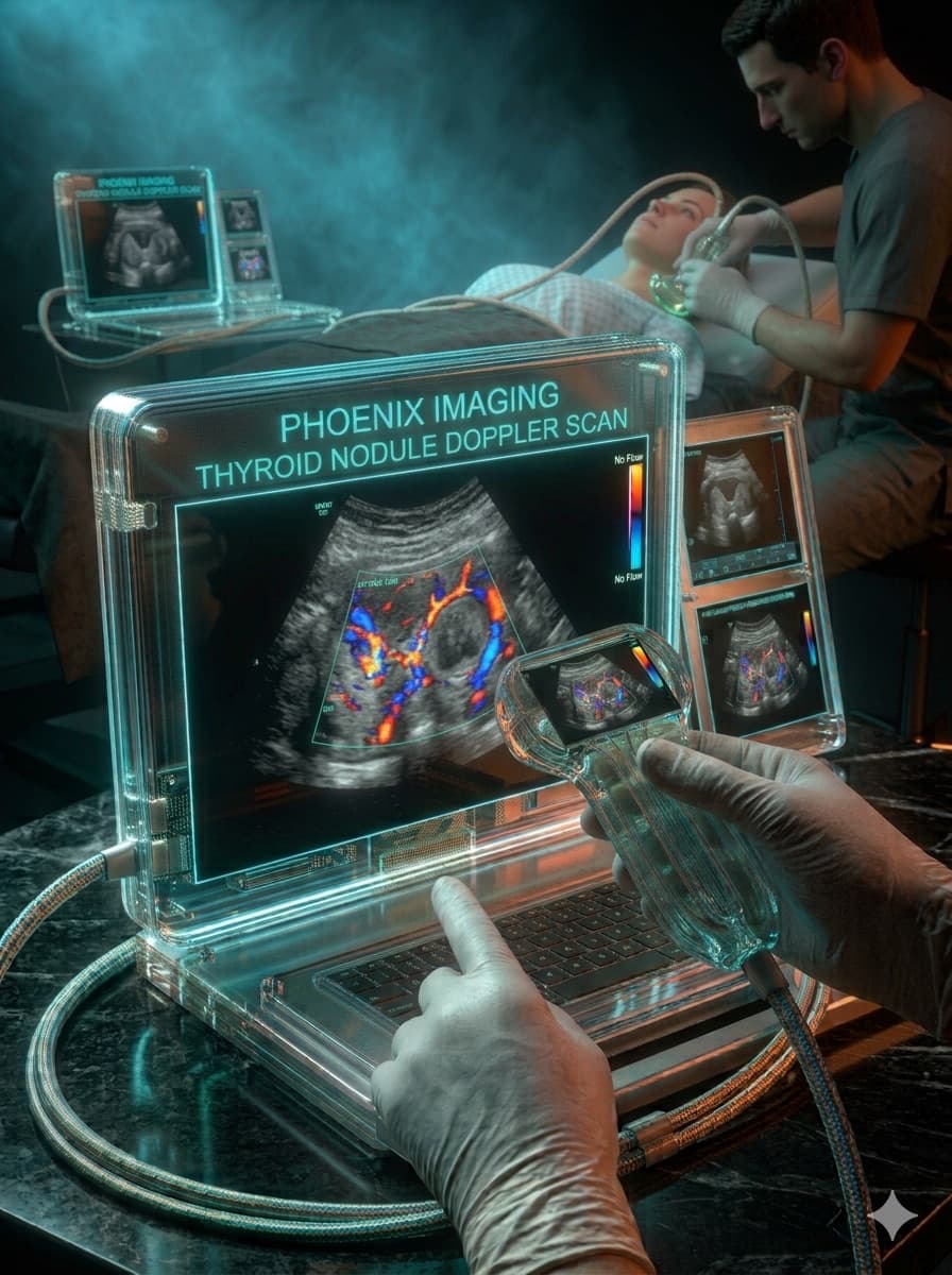
Patient, female, 42 years old, asymptomatic.
Incidental finding of a cervical nodule on routine physical exam, referred for thyroid ultrasound.
High-frequency linear transducer, B-mode assessment in transverse and longitudinal planes, with color Doppler.
Solid, hypoechoic nodule with irregular margins, taller than wide, measuring 1.2 cm in the right lobe, without macrocalcifications.
Predominantly central vascularity on color Doppler.
Atypical colloid nodule · Follicular adenoma · Papillary carcinoma
Nodule classified as TI-RADS 4, with indication for ultrasound-guided fine-needle aspiration.
Key points
- Irregular margins and a taller-than-wide shape carry the most weight in risk stratification.
- The TI-RADS classification guides the choice between follow-up and biopsy — it does not replace clinical judgment.
- Size alone does not define malignancy; morphology is the central criterion.
Illustrative, anonymized case for educational purposes only. Does not represent a real patient and does not replace individualized medical evaluation.
Want to see more cases like this?
Join the Ultrassom 360° VIP list and get commented clinical cases, free lessons, and news before everyone else.