Hand paresthesia — assessment of the median nerve at the carpal tunnel
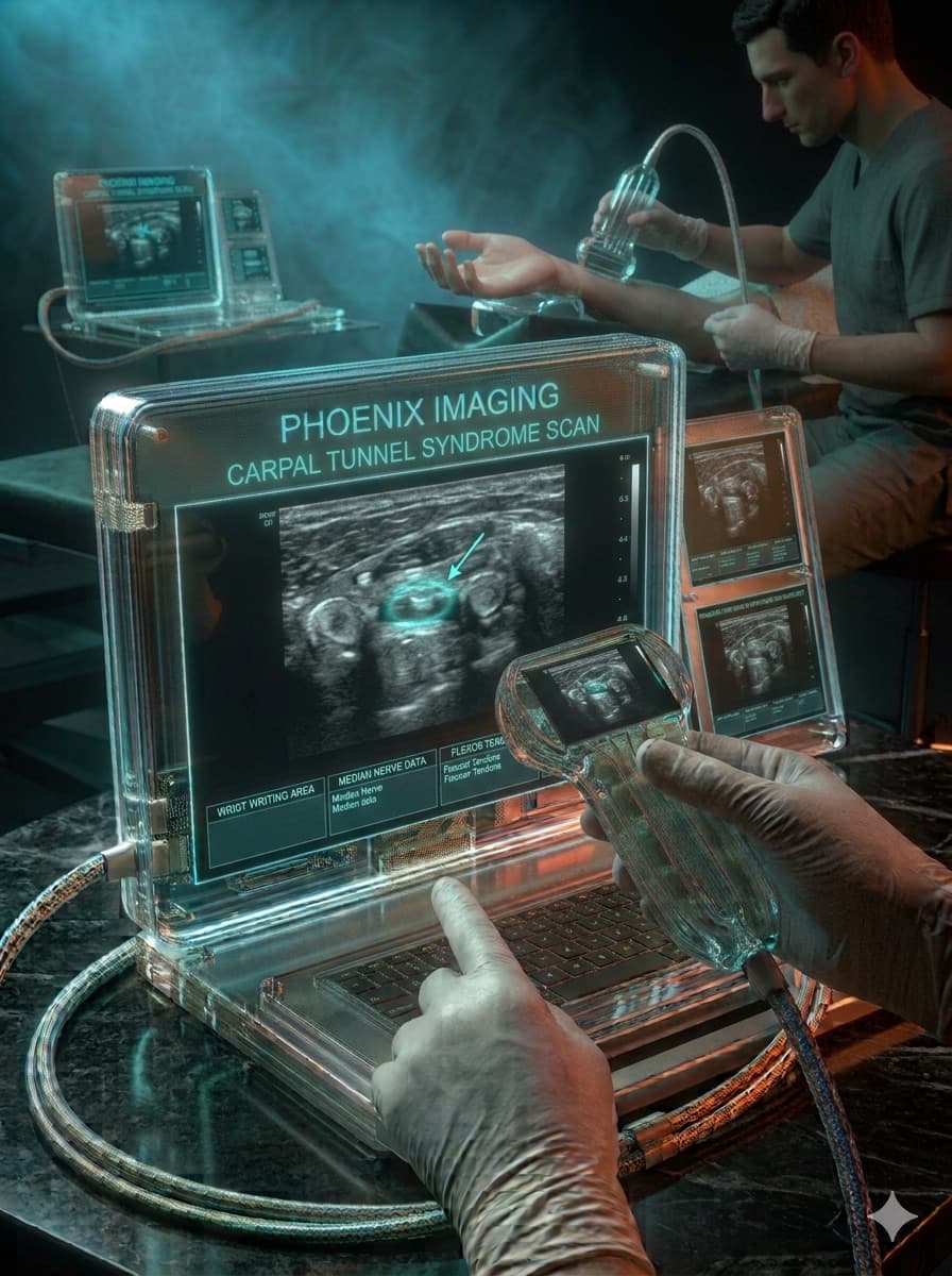
Patient, female, 48 years old, data entry worker, bilateral nocturnal paresthesia.
Paresthesia in the first, second, and third digits, worse at night, bilateral.
High-frequency linear transducer, cross-sectional area of the median nerve measured at the carpal tunnel inlet, with dynamic assessment during finger flexion-extension.
Median nerve cross-sectional area of 14 mm² on the right and 13 mm² on the left, with distal flattening and bowing of the flexor retinaculum.
Carpal tunnel syndrome · Peripheral neuropathy of another etiology · Associated flexor tenosynovitis
Findings consistent with bilateral carpal tunnel syndrome of moderate severity.
Key points
- A cross-sectional area above 10 mm² is the most widely used ultrasound criterion.
- Distal flattening and retinaculum bowing reinforce the dynamic diagnosis.
- Bilateral assessment is recommended even when symptoms are predominantly unilateral.
Illustrative, anonymized case for educational purposes only. Does not represent a real patient and does not replace individualized medical evaluation.
Want to learn to work up cases like this?
Get the free lesson "5 mistakes that make you miss important findings in musculoskeletal ultrasound," with Dr. Andrey Garcez.